Detection of X-rays is very specific because high-energy photons interact differently with the matter. High-energy photons can travel thousands of feet in the air and easily pass through various materials. Moreover, high-energy photons can ionize atoms indirectly and directly (despite they are electrically neutral) through the photoelectric effect and the Compton effect. But secondary (indirect) ionization is much more significant.
To describe the principles of detecting high-energy photons, we must understand the interaction of radiation with matter. Each type of particle interacts differently; therefore, we must describe interactions of high-energy photons (radiation as a flow of these rays) separately.
See also: X-Rays
Interaction of X-rays with Matter
Although many possible interactions are known, there are three key interaction mechanisms with the matter. The strength of these interactions depends on the X-rays’ energy and the material’s elemental composition. Still, not much on chemical properties, since the X-ray photon energy is much higher than chemical binding energies. The photoelectric absorption dominates at low-energies of X-rays, while Compton scattering dominates at higher energies.
The photon is completely absorbed in the photoelectric effect, while only partial energy is deposited in any given Compton scattering. The probability of photoelectric absorption (dominates at lower X-rays energies) per unit mass is approximately proportional to:
τ(photoelectric) = constant x ZN/E3.5
where Z is the atomic number, and the exponent n varies between 4 and 5. E is the energy of the incident photon. The probability of Compton scattering per one interaction with an atom increases linearly with atomic number Z because it depends on the number of electrons available for scattering in the target atom.
Detectors of X-Rays
Detectors may also be categorized according to sensitive materials and methods that can be utilized to make a measurement:
Detection of X-Rays using Ionization Chamber
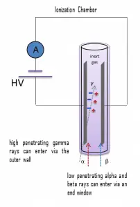
Gamma rays have very little trouble penetrating the metal walls of the chamber. Therefore, ionization chambers may be used to detect gamma radiation and X-rays, collectively known as photons, and for this, the windowless tube is used. Ionization chambers have a good uniform response to radiation over a wide range of energies and are the preferred means of measuring high levels of gamma radiation. Some problems are caused by alpha particles being more ionizing than beta particles, and gamma rays, so more current is produced in the ionization chamber region by alpha than beta and gamma. Gamma rays deposit a significantly lower amount of energy into the detector than other particles.
Detection of X-Rays using Geiger Counter

Geiger counter can detect ionizing radiation such as alpha and beta particles, neutrons, X-rays, and gamma rays using the ionization effect produced in a Geiger–Müller tube, which gives its name to the instrument. The voltage of the detector is adjusted so that the conditions correspond to the Geiger-Mueller region.
The high amplification factor of the Geiger counter is the major advantage over the ionization chamber. The Geiger counter is a much more sensitive device than other chambers. This is often used to detect low-level gamma rays and beta particles.
Windowless type
Gamma rays have very little trouble penetrating the metal walls of the chamber. Therefore, Geiger counters may be used to detect gamma radiation and X-rays (thin-walled tubes) collectively known as photons, and for this, the windowless tube is used.
- A thick-walled tube is used for gamma radiation detection above energies of about 25 KeV, and this type generally has an overall wall thickness of about 1-2 mm of chrome steel.
- A thin-walled tube is used for low-energy photons (X-rays or gamma rays) and high-energy beta particles. The transition from thin-walled to thick-walled design takes place at the 300–400 keV energy levels. Above these levels, thick walled designs are used, and the direct gas ionization effect is predominant beneath these levels.
Detection of X-rays using Scintillation Counter
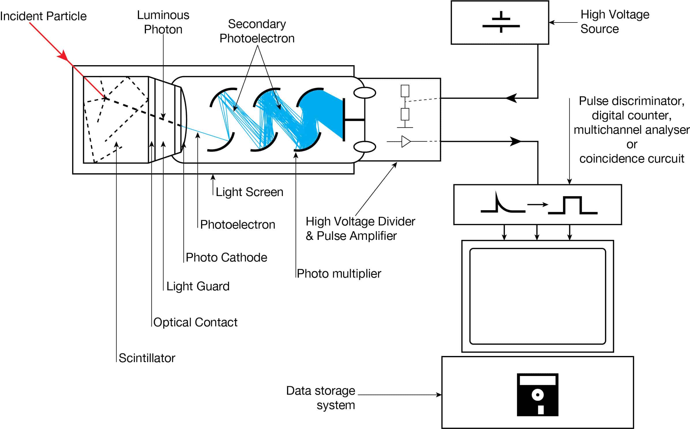
Scintillation counters are used to measure radiation in various applications, including hand-held radiation survey meters, personnel and environmental monitoring for radioactive contamination, medical imaging, radiometric assay, nuclear security, and nuclear plant safety. They are widely used because they can be made inexpensively yet with good efficiency and can measure both the intensity and the energy of incident radiation.
Scintillation counters can detect alpha, beta, X-rays, and gamma radiation and can also be used to detect neutrons. For these purposes, different scintillators are used.
- X-Rays. High-Z materials are best suited as scintillators for the detection of gamma rays. NaI(Tl) (thallium-doped sodium iodide) is the most widely used scintillation material. The iodine provides most of the stopping power in sodium iodide (since it has a high Z = 53). These crystalline scintillators are characterized by high density, high atomic number, and pulse decay times of approximately 1 microsecond (~ 10-6 sec). Scintillation in inorganic crystals is typically slower than in organic ones. They exhibit high efficiency for the detection of gamma rays and are capable of handling high count rates. Inorganic crystals can be cut to small sizes and arranged in an array configuration to provide position sensitivity. This feature is widely used in medical imaging to detect X-rays or gamma rays. Inorganic scintillators are better at detecting gamma rays and X-rays. This is due to their high density and atomic number, which gives a high electron density.
Detection of X-Rays using Semiconductors – HPGe Detectors
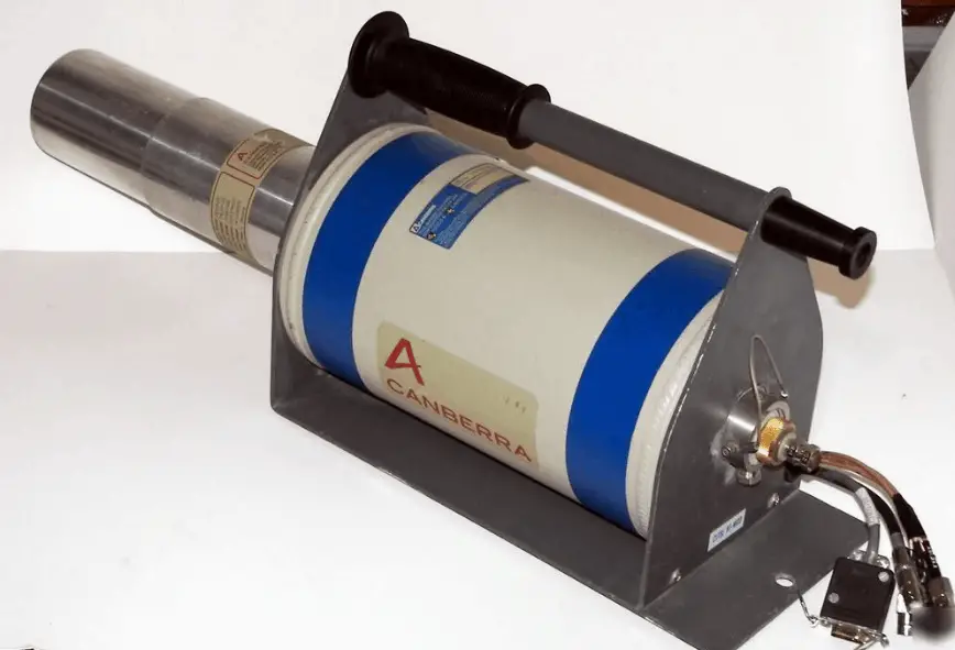
High-purity germanium detectors (HPGe detectors) are the best solution for precise gamma and x-ray spectroscopy.
As was written, the study and analysis of gamma-ray spectra for scientific and technical use are called gamma spectroscopy. Gamma-ray spectrometers are the instruments that observe and collect such data. A gamma-ray spectrometer (GRS) is a sophisticated device for measuring the energy distribution of gamma radiation. For the measurement of gamma rays above several hundred keV, there are two detector categories of major importance, inorganic scintillators such as NaI(Tl) and semiconductor detectors. If a perfect energy resolution is required, we have to use a germanium-based detector, such as the HPGe detector. Germanium-based semiconductor detectors are most commonly used where a very good energy resolution is required, especially for gamma spectroscopy as well as x-ray spectroscopy. In gamma spectroscopy, germanium is preferred due to its atomic number being much higher than silicon, increasing the probability of gamma-ray interaction. Moreover, germanium has lower average energy necessary to create an electron-hole pair, which is 3.6 eV for silicon and 2.9 eV for germanium. This also provides the latter with a better resolution in energy. The FWHM (full width at half maximum) for germanium detectors is an energy function. For a 1.3 MeV photon, the FWHM is 2.1 keV, which is very low.